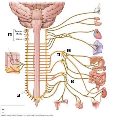

Two upward arrows branch from the neuron’s dendrites. The second image is a neuron containing dendrites, axon, and axon terminals labeled temperature receptors. Two downward arrows point to an aorta of a heart, and one arrow points down to the hypothalamus and pituitary gland in the brain. Three arrows branch out from the cell body of the neuron. The first image is a neuron containing a cell body, axon, and axon terminals labeled chemoreceptors and text listed as follows Blood C O subscript 2, O subscript 2 blood pH blood osmotic pressure and blood glucose levels. Visceral receptorsĪn image shows a multipart illustration of the visceral receptors. The autonomic control centers then send action potentials down motor pathways to inhibit or excite ANS effectors. The ANS responds to unconsciously perceived visceral sensations. Text at the bottom of the screen reads The autonomic nervous system ANS is defined primarily as a set of motor pathways to regulate visceral activity. Each of the aforementioned images links to a more detailed screen. The motor pathway neuron’s axon terminals connect with the second circle, which contains the ball-and-stick model of A C h. The motor pathway branch that leads into visceral effectors connects with the A C h and N E circle.

The first circle is divided into two halves that contain the ball-and-stick models of A C h and N E. The third image is the neurotransmitters, which have two circles. The second image is a box containing smooth muscles, heart, and adrenal gland labeled visceral effectors. Inside the circular structure, a neuron’s cell body is present, which continues as axon axon terminals lead to the second image.

The neuron at the bottom, labeled motor pathway, has dendrites emerging from the center to continue as axon, and branches out into a circular structure containing the axon terminals. The center of the spinal cord is labeled control center. The neuron at the top, labeled visceral receptor, has dendrites emerging from the center of the spinal cord to continue as axon and end as axon terminals. Two neurons emerge from the center of the spinal cord. The first image is a cross-section of a spinal cord with receptors and pathways labeled. Anatomy Overview: Nervous System: Organization of the ANS Autonomic nervous systemĪn image shows a three-part illustration of the autonomic nervous system.


 0 kommentar(er)
